
August 2017 Pulmonary Case of the Month
Lewis J. Wesselius, MD
Department of Pulmonary Medicine
Mayo Clinic Arizona
Scottsdale, AZ USA
History of Present Illness
The patient is a 60-year-old woman with dyspnea on exertion when she had a pulmonary embolism following knee surgery 3 years earlier. She smoked 1 pack per day for the past 40 years. She was seen at another hospital and had pulmonary function testing which showed only a DLco which was 66% of predicted. Serologic studies were negative for a rheumatologic disorder. A CT scan was also performed (Figure 1).
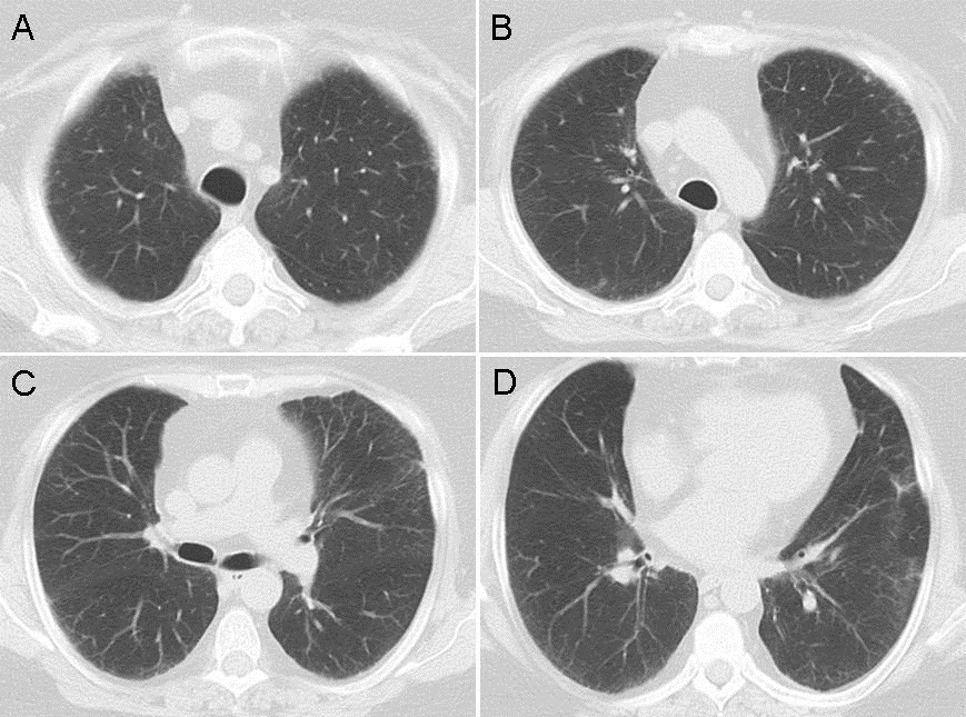
Figure 1. Representative images from thoracic CT scan in lung windows.
The CT scan was interpreted as showing a few small nodules and possible very early interstitial lung disease.
Which of the following are true? (Click on the correct answer to proceed to the second of five pages)
- A pulmonary embolism can reduce the DLco
- Her CT scan is characteristic of Langerhans cell histiocytosis
- Smoking can reduce the DLco
- 1 and 3
- All of the above
Cite as: Wesselius LJ. August 2017 pulmonary case of the month. Southwest J Pulm Crit Care. 2017;15(2):54-60. doi: https://doi.org/10.13175/swjpcc096-17 PDF
 Post a Comment
Reader Comments