
Thursday
Nov052015
November 2015 Imaging Case of the Month
Michael B. Gotway, MD
Department of Radiology
Mayo Clinic Arizona
Scottsdale, AZ
Clinical History: A 48-year-old non-smoking woman with a history of hysterectomy and right oophorectomy, and cholecystectomy, otherwise previously healthy, presented with right-sided chest pain. A frontal chest radiograph (Figure 1) was performed.
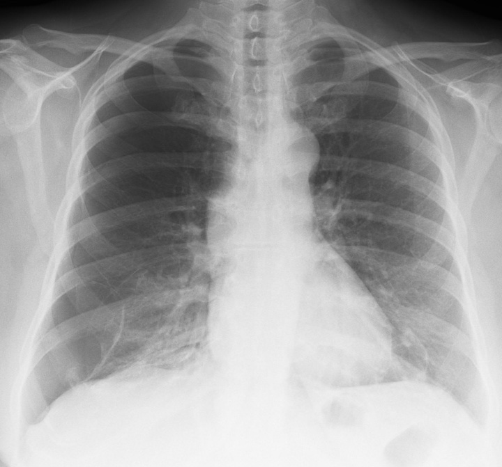
Figure 1. Frontal chest radiograph.
Which of the following statements regarding the chest radiograph is most accurate? (Click on the correct answer to proceed to the second of eight panels)
Cite as: Gotway MB. November 2015 imaging case of the month. Southwest J Pulm Crit Care. 2015;11(5):218-25. doi: http://dx.doi.org/10.13175/swjpcc140-15 PDF
 Post a Comment
Reader Comments