Medical Image of the Week: Aortic Ring
 Wednesday, October 15, 2014 at 8:00AM
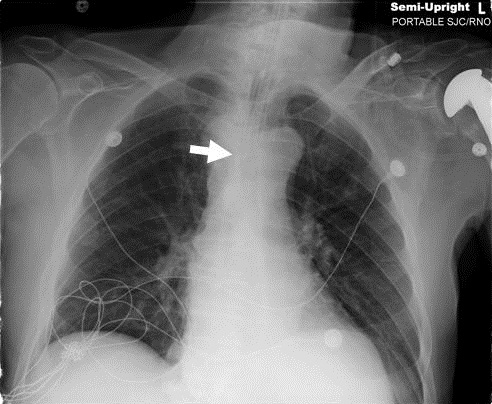
Figure 1. Post-intubation chest radiograph showing a widened right paratracheal stripe (arrow).
A 78 year old man presented with altered mental status and was found to have an intraventricular hemorrhage. He was intubated for airway protection. On the post-intubation chest radiograph (Figure 1), the patient was noted to have a widening of the right paratracheal stripe.
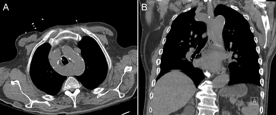
A CT chest (Figure 2) was obtained to characterize this finding and revealed an aortic ring which encircles the trachea and esophagus.

Figure 2. Axial (Panel A) and coronal (Panel B) of thoracic CT soft tissue windows showing the aortic ring.
Vascular rings are uncommon congenital abnormalities, accounting for approximately 1% of congenital heart disease. Complete vascular rings can occur with a right aortic arch with a ligamentum arteriosum or with a double aortic arch, such as with our patient (1). This ring can cause airway compression, stridor, esophageal compression, or no symptoms at all. As the embryo develops, the left fourth pharyngeal arch normally persists to become the aortic arch while the right fourth pharyngeal arch regresses. If both fourth pharyngeal arches persist, a right aortic arch can form and surround the trachea and esophagus (2).
Candy Wong MD1, Tammer Elaini MD2, Soyoung Park MD2, Scott Rosen MD2, and Sai Parthasarathy MD1
1Division of Pulmonary, Allergy, Critical Care, and Sleep Medicine. Department of Medicine
2Department of Medicine
University of Arizona
Tucson, AZ
References
-
Turner A, Gavel G, Coutts J. Vascular rings-presentation, investigation and outcome. Eur J Pediatr. 2005;164(5):266-70. [CrossRef] [PubMed]
-
Vatish J, McCarthy R, Perris R. A double aortic arch presenting in the 7th decade of life. J Surg Case Rep. 2013.(10):pii:rjt081. [CrossRef] [PubMed]
Reference as: Wong C, Elaini T, Park S, Rosen S, Parthasarathy S. Medical image of the week: aortic ring. Southwest J Pulm Crit Care. 2014;9(4):121-2. doi: http://dx.doi.org/10.13175/swjpcc130-14 PDF

Reader Comments