Search Journal-type in search term and press enter
Southwest Pulmonary and Critical Care Fellowships
In Memoriam

Imaging

Last 50 Imaging Postings

(Most recent listed first. Click on title to be directed to the manuscript.)

February 2025 Medical Image of the Month: Unexpected Complications of
   Transjugular Intrahepatic Portosystemic Shunt (TIPS) 
February 2025 Imaging Case of the Month: A Wolf in Sheep’s Clothing
January 2025 Medical Image of the Month: Psoriasis with Pulmonary
   Involvement
December 2024 Medical Image of the Month: An Endobronchial Tumor
November 2024 Medical Image of the Month: A Case of Short Telomeres
November 2024 Imaging Case of the Month: A Recurring Issue
October 2024 Medical Image of the Month: Lofgren syndrome with Erythema
   Nodosum
September 2024 Medical Image of the Month: A Curious Case of Nasal
   Congestion
August 2024 Image of the Month: Lymphomatoid Granulomatosis
August 2024 Imaging Case of the Month: An Unexplained Pleural Effusion
July 2024 Medical Image of the Month: Vocal Cord Paralysis on PET-CT 
June 2024 Medical Image of the Month: A 76-year-old Man Presenting with
   Acute Hoarseness
May 2024 Medical Image of the Month: Hereditary Hemorrhagic
   Telangiectasia in a Patient on Veno-Arterial Extra-Corporeal Membrane
   Oxygenation
May 2024 Imaging Case of the Month: Nothing Is Guaranteed
April 2024 Medical Image of the Month: Wind Instruments Player Exhibiting
   Exceptional Pulmonary Function
March 2024 Medical Image of the Month: Sputum Cytology in Patients with
   Suspected Lung Malignancy Presenting with Acute Hypoxic Respiratory
   Failure
February 2024 Medical Image of the Month: Pulmonary Alveolar Proteinosis
   in Myelodysplastic Syndrome
February 2024 Imaging Case of the Month: Connecting Some Unusual Dots
January 2024 Medical Image of the Month: Polyangiitis Overlap Syndrome
   (POS) Mimicking Fungal Pneumonia 
December 2023 Medical Image of the Month: Metastatic Pulmonary
   Calcifications in End-Stage Renal Disease 
November 2023 Medical Image of the Month: Obstructive Uropathy
   Extremis
November 2023 Imaging Case of the Month: A Crazy Association
October 2023 Medical Image of the Month: Swyer-James-MacLeod
   Syndrome
September 2023 Medical Image of the Month: Aspergillus Presenting as a
   Pulmonary Nodule in an Immunocompetent Patient
August 2023 Medical Image of the Month: Cannonball Metastases from
   Metastatic Melanoma
August 2023 Imaging Case of the Month: Chew Your Food Carefully
July 2023 Medical Image of the Month: Primary Tracheal Lymphoma
June 2023 Medical Image of the Month: Solitary Fibrous Tumor of the Pleura
May 2023 Medical Image of the Month: Methamphetamine Inhalation
Leading to Cavitary Pneumonia and Pleural Complications
April 2023 Medical Image of the Month: Atrial Myxoma in the setting of
   Raynaud’s Phenomenon: Early Echocardiography and Management of
   Thrombotic Disease
April 2023 Imaging Case of the Month: Large Impact from a Small Lesion
March 2023 Medical Image of the Month: Spontaneous Pneumomediastinum
   as a Complication of Marijuana Smoking Due to Müller's Maneuvers
February 2023 Medical Image of the Month: Reversed Halo Sign in the
   Setting of a Neutropenic Patient with Angioinvasive Pulmonary
   Zygomycosis
January 2023 Medical Image of the Month: Abnormal Sleep Study and PFT
   with Supine Challenge Related to Idiopathic Hemidiaphragmatic Paralysis
December 2022 Medical Image of the Month: Bronchoesophageal Fistula in
   the Setting of Pulmonary Actinomycosis
November 2022 Medical Image of the Month: COVID-19 Infection
   Presenting as Spontaneous Subcapsular Hematoma of the Kidney
November 2022 Imaging Case of the Month: Out of Place in the Thorax
October 2022 Medical Image of the Month: Infected Dasatinib Induced
   Chylothorax-The First Reported Case 
September 2022 Medical Image of the Month: Epiglottic Calcification
Medical Image of the Month: An Unexpected Cause of Chronic Cough
August 2022 Imaging Case of the Month: It’s All About Location
July 2022 Medical Image of the Month: Pulmonary Nodule in the
   Setting of Pyoderma Gangrenosum (PG) 
June 2022 Medical Image of the Month: A Hard Image to Swallow
May 2022 Medical Image of the Month: Pectus Excavatum
May 2022 Imaging Case of the Month: Asymmetric Apical Opacity–
   Diagnostic Considerations
April 2022 Medical Image of the Month: COVID Pericarditis
March 2022 Medical Image of the Month: Pulmonary Nodules in the
   Setting of Diffuse Idiopathic Pulmonary NeuroEndocrine Cell Hyperplasia
   (DIPNECH) 
February 2022 Medical Image of the Month: Multifocal Micronodular
   Pneumocyte Hyperplasia in the Setting of Tuberous Sclerosis
February 2022 Imaging Case of the Month: Between A Rock and a
   Hard Place
January 2022 Medical Image of the Month: Bronchial Obstruction
   Due to Pledget in Airway Following Foregut Cyst Resection
December 2021 Medical Image of the Month: Aspirated Dental Implant
Medical Image of the Month: Cavitating Pseudomonas
   aeruginosa Pneumonia
November 2021 Imaging Case of the Month: Let’s Not Dance
   the Twist
Medical Image of the Month: COVID-19-Associated Pulmonary
   Aspergillosis in a Post-Liver Transplant Patient
Medical Image of the Month: Stercoral Colitis
Medical Image of the Month: Bleomycin-Induced Pulmonary Fibrosis
   in a Patient with Lymphoma
August 2021 Imaging Case of the Month: Unilateral Peripheral Lung
   Opacity


For complete imaging listings click here

Those who care for patients with pulmonary, critical care or sleep disorders rely heavily on chest radiology and pathology to determine diagnoses. The Southwest Journal of Pulmonary, Critical Care & Sleep publishes case-based articles with characteristic chest imaging and related pathology. The editor of this section will oversee and coordinate the publication of a core of the most important chest imaging topics. In doing so, they encourage the submission of unsolicited manuscripts. It cannot be overemphasized that both radiologic and pathologic images must be of excellent quality. As a rule, 600 DPI is sufficient for radiographic and pathologic images. Taking pictures of plain chest radiographs and CT scans with a digital camera is strongly discouraged. The figures should be cited in the text and numbered consecutively. The stain used for pathology specimens and magnification should be mentioned in the figure legend. Those who care for patients with pulmonary, critical care or sleep disorders rely heavily on chest radiology and pathology to determine diagnoses. The Southwest Journal of Pulmonary, Critical Care & Sleep publishes case-based articles with characteristic chest imaging and related pathology. The editor of this section will oversee and coordinate the publication of a core of the most important chest imaging topics. In doing so, they encourage the submission of unsolicited manuscripts. It cannot be overemphasized that both radiologic and pathologic images must be of excellent quality. As a rule, 600 DPI is sufficient for radiographic and pathologic images. Taking pictures of plain chest radiographs and CT scans with a digital camera is strongly discouraged. The figures should be cited in the text and numbered consecutively. The stain used for pathology specimens and magnification should be mentioned in the figure legend.

-------------------------------------------------------------------------------------------  

Sunday
Feb022025

February 2025 Medical Image of the Month: Unexpected Complications of Transjugular Intrahepatic Portosystemic Shunt (TIPS)

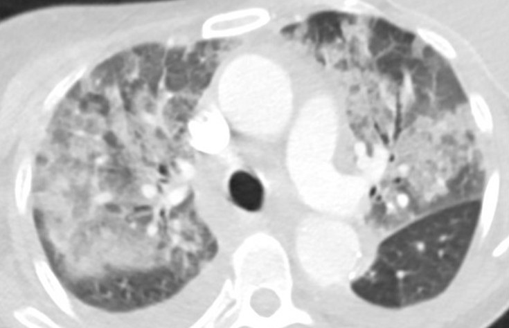
Figure 1. Axial “lung windows” reconstruction from a contrast-enhanced CT angiogram demonstrating mixed interstitial and airspace opacities consisting of smooth septal lines with patchy superimposed consolidation and ground glass with a central distribution (peripheral sparing). There are also small layering pleural effusions. Findings are nonspecific but would be considered consistent with “batwing edema” in the setting of severe alveolar edema given the clinical context. To view Figure 1 in a separate, enlarged window click here.

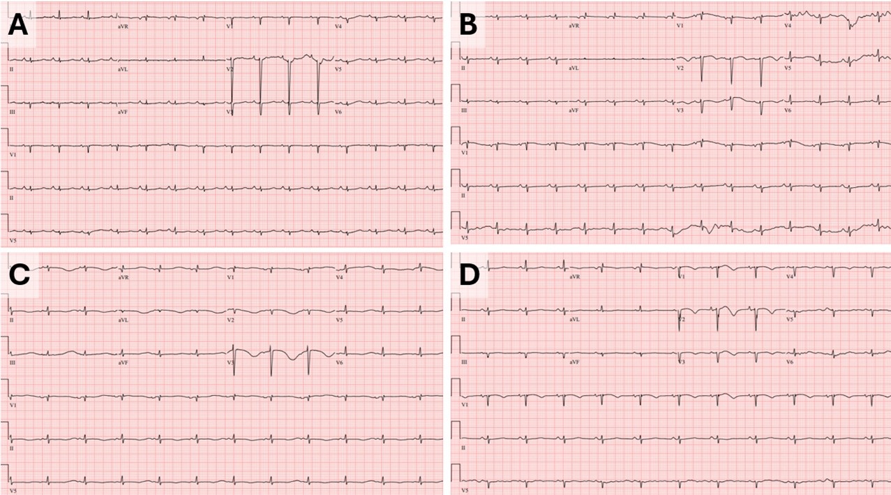
Figure 2. Baseline ECG (A) performed 2 months prior to tips procedure: HR 90bpm, QTC 513ms. Admission ECG (B): HR 88, QTC 568ms. ECG a few hours prior to episode of V-tach (C): HR 70, QTC 726ms. ECG prior to extubation (D): HR 68, QTc 523ms. To view Figure 2 in a separate, enlarged window click here.

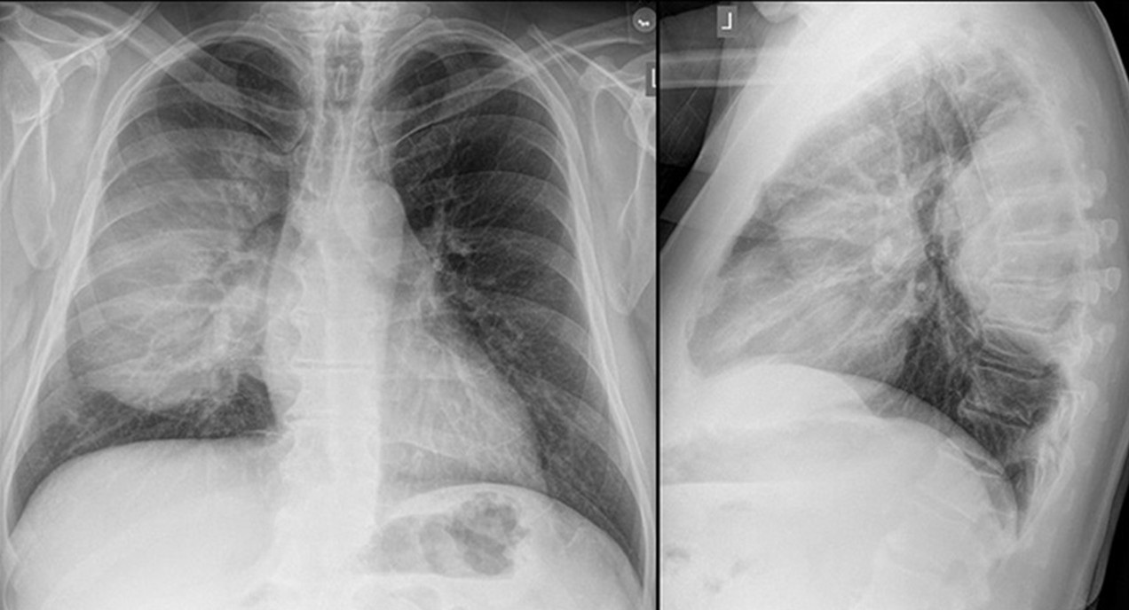
A 55-year-old woman with cirrhosis secondary to alcohol use disorder presented to the emergency department with worsening shortness of breath, orthopnea, and two recent syncopal episodes with seizure-like activity. She had undergone a transjugular intrahepatic portosystemic shunt (TIPS) procedure for refractory ascites one month prior. On arrival, she was hemodynamically stable but tachypneic (respiratory rate 22 bpm) with an SpO2 of 90% on room air. Her jugular venous pressure (JVP) was elevated. Chest X-ray showed bilateral pulmonary infiltrates. CT angiography (Figure 1) revealed bilateral patchy airspace disease with a central distribution and pleural effusions, suggesting alveolar edema. Abdominal imaging confirmed liver disease, ascites, and a TIPS. Laboratory tests revealed negative respiratory viral and SARS-CoV-2 PCR tests, ammonia was normal, BNP of 427 pg/mL, normal troponins, sodium 138 mmol/L, potassium 3.7 mmol/L, and procalcitonin 0.23 ng/mL. Blood and sputum cultures were collected. An electrocardiogram (ECG, figure-2B) showed sinus rhythm with QTc of 568 ms (baseline EKG Figure-2A). The patient was admitted to the step-down unit and started on broad-spectrum antibiotics for presumed pneumonia.

The following day she continued to have shortness of breath and also developed nausea, for which ondansetron 4 mg IV was ordered. She also received hydroxyzine which is a regular home medication. Shortly after, she developed supraventricular tachycardia (SVT) at 200 bpm, unresponsive to adenosine but controlled with 5 mg of metoprolol. Two hours later, she developed polymorphic ventricular tachycardia (V-tach) which led to cardiac arrest. After two minutes of cardiopulmonary resuscitation (CPR), spontaneous circulation was restored, and the patient was intubated and transferred to the ICU. Repeat ECG (Figure-2C) following cardiac arrest showed a prolonged QTc interval of 726 ms. In the ICU, she was sedated with a midazolam drip and diuresed with furosemide. She received magnesium and potassium replacement. Potential QT-prolonging medications were discontinued, and she was started on metoprolol 12.5 mg twice daily. Her QTc improved to 523 ms (figure-2D). Chest X-ray showed significant improvement over 24 hours, and a repeat echocardiogram showed normal ejection fraction. The patient was extubated and transferred back to the medical floor for further care. A PharmD team reviewed her medications before discharge, and she was educated on avoiding QT-prolonging drugs.

The liver is crucial in drug metabolism, primarily through the cytochrome P450 enzyme system, accounting for 40-50% of its activity. In cirrhotic patients, this activity is markedly reduced, impairing drug clearance and leading to the accumulation of QT-prolonging drugs. Additionally, cirrhotic patients have diminished intestinal CYP3A4 activity (approximately 30-40% of normal) (1). The QT interval is often prolonged in patients with both non-cirrhotic and cirrhotic portal hypertension, and portal decompression through TIPS exacerbates this abnormality (2). Furthermore, the intravenous route translates to faster drug bioavailability and higher peak blood concentrations compared to oral administration. Although this phenomenon has not been extensively studied in cirrhotic patients, it has been evaluated in emergency department patients receiving intravenous ondansetron (3). The presence of a TIPS may prolong the drug half-life, potentially increasing the risk of severe arrhythmias, such as torsades de pointes. This might explain the onset and the timing of life-threatening arrhythmias observed in our patient following the administration of intravenous ondansetron and oral hydroxyzine. In retrospect, the syncopal episodes observed at home in our case likely indicated episodes of ventricular tachycardia. Our patient had no prior history of similar events before undergoing the TIPS procedure. The combination of impaired liver function, metabolic disturbances, and the addition of a TIPS creates a "perfect storm" for life-threatening arrhythmias, particularly in those who are already on QT-prolonging medication.

Late onset pulmonary edema is another atypical presentation our case. Following a TIPS procedure, JVP may not reliably reflect the patient's true fluid status or heart function. An improvement in central hypovolemia could be attributed to an increase in thoracic blood volume, encompassing both central venous and arterial components. This is further reinforced by an increase in preload and a simultaneous decrease in afterload (4). Chronic liver disease with portal hypertension is associated with increased cardiac output, reduced peripheral vascular resistance, and normal cardiac filling pressures. Studies by Azoulay et al. (5) suggest that after the creation of a portosystemic shunt during TIPS, cardiac filling pressures rise, and a hyperdynamic cardiac state may persist for up to a month, increasing the risk of pulmonary edema. While pulmonary edema is typically an early complication post-TIPS (6), our case presented a delayed onset, which, to our knowledge, has not been previously described.

This case highlights the importance of recognizing potential complications following TIPS, particularly QTc prolongation and delayed pulmonary edema. The risk of severe QTc prolongation is heightened when patients are exposed to QT-prolonging medications. Healthcare providers should be aware of these risks and closely monitor for such life-threatening complications post TIPS.

Abdulmonam Ali, MD 

SSM Health

Mount Vernon, IL USA

References

  1. Vuppalanchi R, Juluri R, Ghabril M, Kim S, Thong N, Gorski JC, Chalasani N, Hall SD. Drug-induced QT prolongation in cirrhotic patients with transjugular intrahepatic portosystemic shunt. J Clin Gastroenterol. 2011 Aug;45(7):638-42. [CrossRef] [PubMed]
  2. Trevisani F, Merli M, Savelli F, Valeriano V, Zambruni A, Riggio O, Caraceni P, Domenicali M, Bernardi M. QT interval in patients with non-cirrhotic portal hypertension and in cirrhotic patients treated with transjugular intrahepatic porto-systemic shunt. J Hepatol. 2003 Apr;38(4):461-7. [CrossRef] [PubMed]
  3. Rezaei Zadeh Rukerd M, Shahrbabaki FR, Movahedi M, Honarmand A, Pourzand P, Mirafzal A. Single intravenous dose ondansetron induces QT prolongation in adult emergency department patients: a prospective observational study. Int J Emerg Med. 2024 Apr 2;17(1):49. [CrossRef] [PubMed]
  4. Busk TM, Bendtsen F, Poulsen JH, et al. Transjugular intrahepatic portosystemic shunt: impact on systemic hemodynamics and renal and cardiac function in patients with cirrhosis. Am J Physiol Gastrointest Liver Physiol. 2018 Feb 1;314(2):G275-G286. [CrossRef] [PubMed]
  5. Azoulay D, Castaing D, Dennison A, Martino W, Eyraud D, Bismuth H. Transjugular intrahepatic portosystemic shunt worsens the hyperdynamic circulatory state of the cirrhotic patient: preliminary report of a prospective study. Hepatology. 1994 Jan;19(1):129-32. [PubMed]
  6. Willoughby PH, Beers RA, Murphy KD. Pulmonary edema after transjugular intrahepatic portosystemic shunt. Anesth Analg. 1996 Apr;82(4):895-6. [CrossRef] [PubMed]
Cite as: Ali A. February 2025 Medical Image of the Month: Unexpected Complications of Transjugular Intrahepatic Portosystemic Shunt (TIPS). Southwest J Pulm Crit Care Sleep. 2025;30(2):26-29. doi: https://doi.org/10.13175/swjpccs001-25 PDF
Saturday
Feb012025

February 2025 Imaging Case of the Month: A Wolf in Sheep’s Clothing

Mathew T. Stib MD and Michael B. Gotway MD

Department of Radiology

Mayo Clinic Arizona

Phoenix, AZ USA

Clinical History: A 72-year-old man presents to his physician with chest pain. The patient awoke 2 weeks earlier with right-sided chest pain, thought to be the result of a pulled muscle. However, the pain did not remit over the next 2 weeks. The patient also complained of a mild cough productive of white sputum and some mild intermittent shortness of breath improving with albuterol. The patient denied fever.

The patient’s past medical history was remarkable for seasonal allergies, especially dust exposure, treated with fluticasone, and occasional wheezing, treated as needed with an albuterol inhaler. The patient was also diabetic and had hypertension and a history of hypothyroidism. The patient’s past surgical history included right knee total arthroplasty and lithotripsy for renal stones. The patient denied allergies, smoking, and illicit drug use. His medications included fluticasone, albuterol, amlodipine, atorvastatin, ramipril, sitagliptin-metformin, levothyroxine, and probiotics.    

The patient’s vital signs included a blood pressure of 121/75 mmHg, a pulse rate of 78/minute, a respiratory rate of 18/minute and a temperature of 36.4°C. Pulse oximetry on room air was 96%. The patient’s weight was 89.kg, his body mass index 28.15 kg/m2. The physical examination was unremarkable. In particular, the breath sounds were normal.

A complete blood count showed a mildly elevated white blood cell count at 11.1 x 10⁹/L (normal, 3.4 – 9.6 x 10⁹/L), with a normal absolute neutrophil count of 3.36  x 109/L (normal, 1.56 – 6.45 x 10⁹/L). His hemoglobin and hematocrit values were mildly decreased at 11.1 gm/dL (normal, 13.5 – 17.5 gm/dL) and 36.3% (normal, 38.8 – 50%). The platelet count was normal at 280 x 109/L (normal, 149 – 375 x 109/L). The patient’s serum chemistries and liver function studies were normal aside from an a mildly decreased serum calcium level of 8.6 mg/dL (normal, 8.8-10.2 mg/dL), an elevated glucose level of 186 mg/dL, (normal, 70-100 mg/dL), and a mildly elevated alkaline phosphatase level of 121 U/L (normal, 45-115 U/L). The urinary drug toxicity screen was negative, and coagulation parameters were normal. The thyroid stimulating hormone level was within the normal range. Frontal and lateral chest radiography (Figure 1) was performed.

Figure 1. Frontal and lateral chest radiography. To open figure 1 in a separate, enlarged window click here.

Which of the following statements regarding this chest radiograph is most accurate? (click on the correct answer to be directed to the second of eleven pages)

  1. The frontal chest radiograph shows no abnormal findings
  2. The frontal chest radiograph shows basal predominant fibrotic abnormalities
  3. The frontal chest radiograph shows large lung volumes with a cystic appearance
  4. The frontal chest radiograph shows a partially circumscribed opacity projected over the right thorax
  5. The frontal chest radiograph shows abnormal mediastinal contours
Cite as: Stib MT, Gotway MB. February 2025 Imaging Case of the Month: A Wolf in Sheep’s Clothing. Southwest J Pulm Crit Care Sleep. 2025;30(2):14-25. doi: https://doi.org/10.13175/swjpccs003-25 PDF