Ultrasound for Critical Care Physicians: Complication of a Distant Malignancy
 Monday, July 4, 2016 at 8:00AM
 S. Cham Sante M.D.1
Michel Boivin M.D.2
Department of Emergency Medicine1
Division of Pulmonary, Critical care and Sleep Medicine2
University of New Mexico School of Medicine
Albuquerque, NM USA
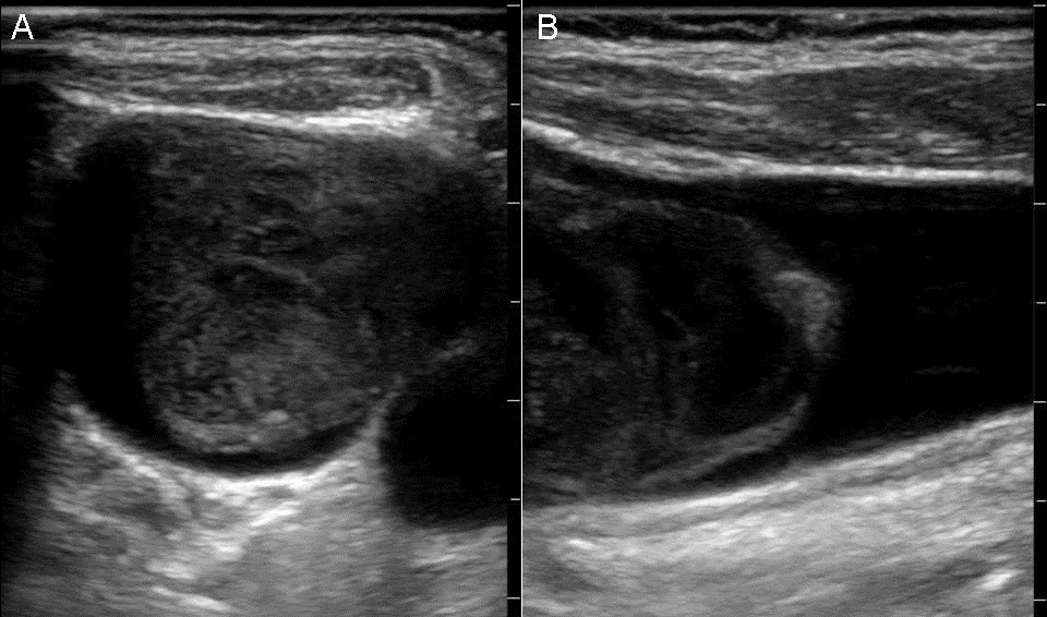
An 82-year-old woman with prior medical history of stage IV colon cancer and chronic obstructive pulmonary disease presented to the medical intensive care unit with newly diagnosed community acquired pneumonia and acute kidney injury. The patient presented with acute onset of shortness of breath, nausea, generalized weakness, bilateral lower extremity swelling and decreased urine output. She was transferred for short term dialysis in the setting of multiple electrolyte abnormalities, including hyperkalemia of 6.4 mmol/l, as well as a creatinine of 6.5 mg/dl. The following imaging of the right internal jugular vein was performed with ultrasound during preparation for placement of a temporary triple lumen hemodialysis catheter.

Figure 1. Panel A: Transverse ultrasound image of the right neck. Panel B: Longitudinal ultrasound image of the right neck, centered on the internal jugular vein.
Based on the above imaging what would be the best location to place the dialysis catheter? (Click on the correct answer for an explanation and discussion)
Cite as: Sante SC, Boivin M. Ultrasound for critical care physicians: complication of a distant malignancy. Southwest J Pulm Crit Care. 2016;13(1):27-9. doi: http://dx.doi.org/10.13175/swjpcc055-16 PDF

Reader Comments