Ultrasound for Critical Care Physicians: The Pleura and the Answers that Lie Within
 Friday, December 4, 2015 at 8:00AM
 Heidi L. Erickson MD
Division of Pulmonary, Critical Care and Occupational Medicine
University of Iowa Hospitals and Clinics
Iowa City, IA
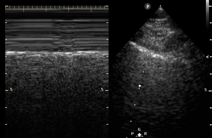
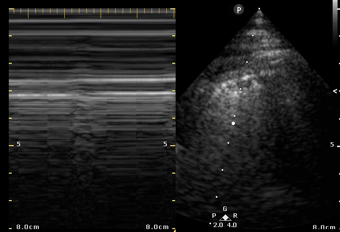
A 67-year-old woman with a 40-pack-year smoking history was admitted to the intensive care unit with acute respiratory failure secondary to adult respiratory distress syndrome (ARDS) in the setting of pneumococcal bacteremia. On admission, she required endotracheal intubation and vasopressor support. She was ventilated using a low tidal volume strategy and was relatively easy to oxygenate with a PEEP of 5 and 40% FiO2. After 48 hours of clinical improvement, the patient developed sudden onset tachypnea and increased peak and plateau airway pressures. A bedside ultrasound was subsequently performed (Figures 1 and 2).

Figure 1. Two- dimensional ultrasound image of the right lung with associated M-mode image.

Figure 2. Two- dimensional ultrasound image of the left lung with associated M-mode image.
What is the cause of this patient’s acute respiratory decompensation and increased airway pressures? (Click on the correct answer for an explanation)
Cite as: Erickson HL. Ultrasound for critical care physicians: the pleura and the answers that lie within. Southwest J Pulm Crit Care. 2015;11(6):260-3. doi: http://dx.doi.org/10.13175/swjpcc149-15 PDF

Reader Comments